Introduction
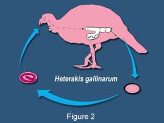
H. gallinaurm is found worldwide in areas where dense plant growth is apparent and it can be protected somewhat from extreme elements (Carron, 2012). The eggs of H. gallinarum can lay in the soil for two weeks before they become infective. As the eggs lay infective in the soil which in some cases can be up to a year (Taylor, Coop & Wall, 2007), it is not uncommon for the earthworm to consume them and become a transport for the host (Taylor, Coop & Wall, 2007). Its life cycle (Fig. 2) then begins again when the fowl then consume these eggs as they forage from the ground (Papini & Cacciuttolo, 2008). The eggs then travel down to the intestine where they then hatch and further develop into adults in the cecum (Shapiro, 2010). The adult H. gallinarum can inhabit the fowl for up to twelve months (Taylor, Coop & Wall, 2007) by surviving off of the contents of the cecum (Carron, 2012). Once the adult female has been fertilized by the adult male and the eggs have fully developed within her uterus, she will then deposit them into the host's feces. Depending upon the species of gallinaceous bird the H. gallinaurm inhabits determines the amount of eggs laid (Carron, 2012). As the fowl defecates the eggs are then passed through with the feces (Shapiro, 2010)
Discussion
H gallinaurm is one of the most frequently diagnosed nematodes in gallinaceous species (Hauck et al., 2012). For example, H gallinaurm has shown to have a prevalence rate of over 84% in the chicken population within the U.S. (Papini & Cacciuttolo, 2008). While observing this prevalence it has shown to be asymptomatic therefore resulting in little threat to the gallinaceous species. However, heavy infestations can result in thickening of the caecal mucosa. Diagnosis is determined by finding the eggs in feces or via necropsy (Taylor, Coop & Wall, 2007).
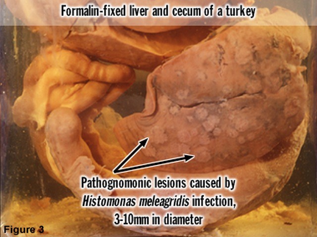
The key importance of this internal parasite is its ability to function as a vector for the protozoan Histomonas meleagridis (Schwarz et al., 2011). The protozoan infects H gallinaurm eggs and becomes highly pathogenic to the host once they ingest the eggs (Brener, Tortelly, Menezes, Muniz-Pereira & Pinto, 2006). According to The Merck Veterinary Manual (2005), the protozoan tends to affect turkeys mostly, but is also found in other gallinaceous species like the chicken. Symptoms start to appear seven to twelve days after being infected with H gallinaurm that include listlessness, drooping wings, unkempt feathers, and yellow droppings. H. meleagridis affects the liver forming focal necrosis leading to developing yellow-green circular depressions while also affecting the cecum resulting in inflammation and ulcers(Fig. 3). A bluish discoloration occurs within the fowl's head resulting in the disease being named "blackhead" (Clinical Information, n.d., para. 3). H. meleagridis can cause a high morbidity rate in chickens (The Merck Veterinary Manual, 2005, para. 3) and a higher mortality rate in turkeys if not treated right away (Clinical Information, n.d., para. 3).
H. gallinaurm is more commonly found in free-rang and organic poultry productions where the gallinaceous species have full access to the outdoors. Infective eggs can remain in the litter if infected fowl are present, so caution should be taken not to reuse any litter as fertilizer (Papini & Cacciuttolo, 2008) as H. gallinaurm can also spread through direct contact. This is especially important when managing a turkey production system because the risk of H. meleagridis infection is greater. Therefore, it is recommended to keep turkeys separated from other domestic poultry (Taylor, Coop & Wall, 2007) because H. meleagridis has been known to cause severe economic loss on turkey production farms (Carron, 2012).
Conclusions
H. gallinaurm, though not considered to be of zoonotic potential to humans (Summary, n.d.) should never be taken lightly as long as H. meleagridis plays a factor. Prevention is possible through good hygiene by means of frequently replacing litter and bedding reducing the amount of eggs left behind in the environment. Poultry production facilities should be advised of the commonness of H. gallinaurm particularly turkey farmers due to the fatal economic impact H. meleagridis can have on their turkey populations. Awareness is power. When we are properly educated about our environment and what lies within it, we become better advocates for promoting overall good health and hygiene.
References
Carron, J. (2012). Heterakis gallinarum. Retrieved from University of Michigan, Animal Diversity Web's Web site: http://animaldiversity.ummz.umich.edu/accounts//.
Taylor, M. A., Coop, R. L., Wall, R. L. (2007). Veterinary Parasitology (3rd ed.). Ames, IA: Blackwell Publishing.
Papini, R. & Cacciuttolo, E. (2008). Observations on the occurrence of Heterakis gallinarum in laying hens kept on soil. Italian Journal of Animal Science, 7 (4), 487-492. Retrieved from http://www.aspajournal.it/index.php/ijas/article/view/404.
Shapiro, L. S. (2010). Pathology and Parasitology for Veterinary Technicians (2nd ed.). New York: Delmar Cengage Learning.
Schwarz, A., Gaulyb M., Abelc, H., Da?b, G., Humburgc, J., Weissd, A., . . . Rautenschleina, S. (2011). Pathobiology of Heterakis gallinarum mono-infection and co-infection with Histomonas meleagridis in layer chickens. Avian Pathology, 40 (3), 277-287. Retrieved from http://www.ncbi.nlm.nih.gov/pubmed/21711187.
Brener, B., Tortelly, R., Menezes, R. C., Muniz-Pereira, L., Pinto, R. M. (2006). Prevalence and pathology of the nematode Heterakis gallinarum, the trematode Paratanaisia bragai, and the protozoan Histomonas meleagridis in the turkey, Meleagris gallopavo. Mem Inst Oswaldo Cruz, Rio de Janeiro, 101 (6), 677-682. Retrieved from http://memorias.ioc.fiocruz.br/101(6)/5614.pdf.
Allen, D., Althouse, G., Ames, T., Andreoni, C., Appel, M., Armstrong, D., . . . Wylie, R. (2005). Histomoniasis: Introduction (Blackhead, Infectious enterohepatitis). Retrieved from http://www.merckvetmanual.com/mvm/index.jsp?cfile=htm/bc/203000.htm.
Heterakis gallinarum. (n.d.). In VetPDA. Retrieved from http://vetpda.ucdavis.edu/parasitolog/Parasite.cfm?ID=75#summary.
Histomonas meleagridis. (n.d.). In VetPDA. Retrieved from http://vetpda.ucdavis.edu/parasitolog/Parasite.cfm?ID=76#clininfo.
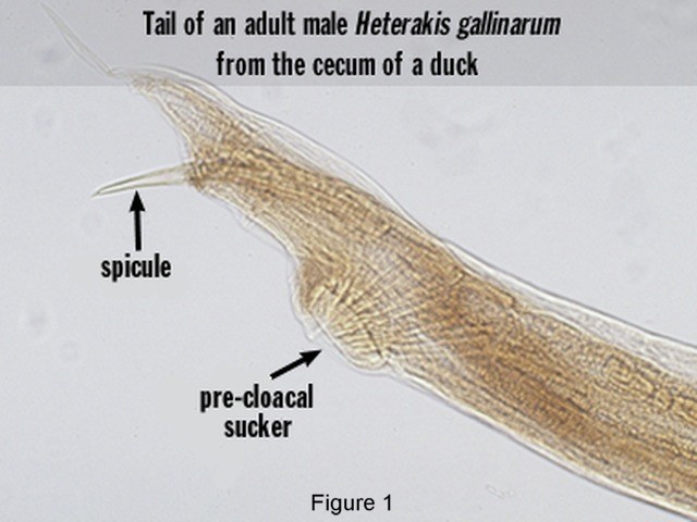
Figure 1: [ Heterakis gallinarum's Tail ]. (n.d.). Retrieved from http://vetpda.ucdavis.edu/parasitolog/Parasite.cfm?ID=75#summary.
Figure 2: [ Heterakis gallinarum's Life Cycle ]. (n.d.). Retrieved from http://vetpda.ucdavis.edu/parasitolog/Parasite.cfm?ID=75#summary.
Figure 3: [ Histomonas meleagridis: Pathogenic Lesions ]. (n.d.). Retrieved from http://vetpda.ucdavis.edu/parasitolog/Parasite.cfm?ID=76#clininfo.